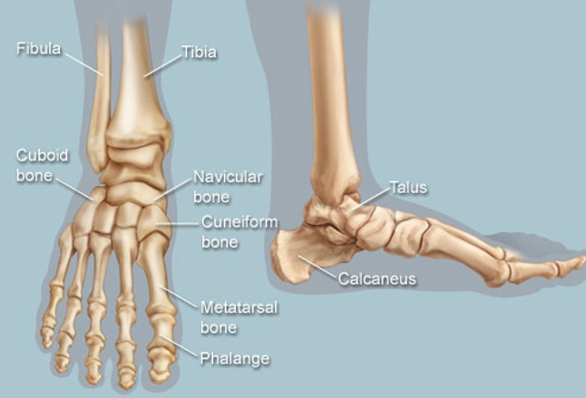
Lower Leg Bone Diagram / Anatomy Of The Foot And Ankle Orthopaedia. Labeled images using 3d reconstructions and an angiographic view. The lower leg contains two major long bones, the tibia and the fibula, which are both very strong skeletal structures. Find stockbilleder af lower leg bone anatomy anterior view i hd og millionvis af andre royaltyfri stockbilleder, illustrationer og vektorer i shutterstocks samling. The foot bones shown in this diagram are the talus, navicular, cuneiform, cuboid, metatarsals and calcaneus. It is thought that tibia refers to both the bone and the musical instrument because flutes were once fashioned from the tibia (of animals).
Hand bone anatomy, wrist anatomy, anatomy bones, human body anatomy, human anatomy and physiology, muscle anatomy, musculoskeletal system, anatomy study, anatomy reference. Two bones make up the lower arm. Most bones (particularly the long bones of the arms and legs — which make up the appendicular skeleton) have a hard outer shell known as bones of the lower extremity. Foot, ankle, & lower leg injuries. The lower leg is the bottom segment of the leg:
Feet Human Anatomy Bones Tendons Ligaments And More from img.webmd.com However, the definition in human anatomy refers only to the section of the lower limb extending from the knee to the ankle, also known as the crus. In humans the head of the fibula is joined to. Your legs are two of your most important body parts. Most bones (particularly the long bones of the arms and legs — which make up the appendicular skeleton) have a hard outer shell known as bones of the lower extremity. 12 photos of the bone anatomy lower leg. Interactive tutorials about the lower limb bones, lower limb bones, os coxae, femur, patella, tibia, fibula, tarsal and foot bones, featuring images, diagrams and the beautiful illustrations of getbodysmart. Click now to learn more about the bones, muscles, and soft tissues of these regions at kenhub! Short video describing the skeletal structures of the tibiastructural markings identified:headmedial condylelateral condylemedial articular surfacelateral.
Why are leg bones thicker than arm bones?
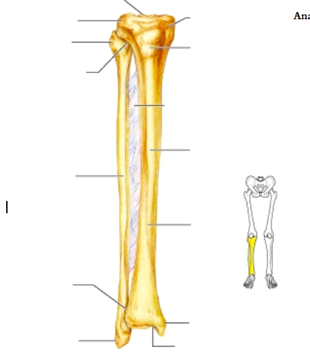
12 photos of the bone anatomy lower leg. Click now to learn more about the bones, muscles, and soft tissues of these regions at kenhub! Labeled images using 3d reconstructions and an angiographic view. The lower leg is the bottom segment of the leg: The lower leg is a major anatomical part of the skeletal system. Here's a diagram with the tibia bone labelled, as well as the fibula, showcasing all their surface here's a leg muscles diagram to give you an overview The part below the knee. Bones give your body structure and enable you to move, but what else is your skeletal system responsible bones prevent you from puddling on the floor in the form of a jellyfish, but what else do they do? Bones of lower leg and foot. Connecting the pelvic girdle to the lower leg is a bone in the thigh area called the femur. Pobierz to zdjęcie infographic diagram of human skeleton lower limb anatomy bone system or leg bone posterior view 3d human anatomy medical diagram educational and human body concept isolated on white background teraz. The thigh bone, or femur, is the large upper leg bone that connects the lower leg bones (knee joint) to the pelvic bone (hip joint). It is thought that tibia refers to both the bone and the musical instrument because flutes were once fashioned from the tibia (of animals).
Connecting the pelvic girdle to the lower leg is a bone in the thigh area called the femur. Bones of lower leg and foot. The human leg, in the general word sense, is the entire lower limb of the human body, including the foot, thigh and even the hip or gluteal region. The two bones beneath your knee that make up your shin are your tibia and fibula. License image the bones of the leg are the femur, tibia, fibula and the foot bones shown in this diagram are the talus, navicular, cuneiform, cuboid, metatarsals and fibula, outer of two bones of the lower leg or hind limb.
Lower Leg Bones Diagram Quizlet from o.quizlet.com Anterior view with primary bones names. Pobierz to zdjęcie infographic diagram of human skeleton lower limb anatomy bone system or leg bone posterior view 3d human anatomy medical diagram educational and human body concept isolated on white background teraz. Two bones make up the lower arm. The lower leg is the bottom segment of the leg: Labeled images using 3d reconstructions and an angiographic view. Your upper and lower leg are connected by a hinge joint. However, the definition in human anatomy refers only to the section of the lower limb extending from the knee to the ankle, also known as the crus. The bones of the leg are the femur, tibia, fibula and patella.
The tibia, or shin bone, spans the lower leg, articulating proximally with the femur and patella at the knee joint, and distally with the tarsal bones, to form the ankle joint.
Foot, ankle, & lower leg injuries. Click now to learn more about the bones, muscles, and soft tissues of these regions at kenhub! It widens and forms two condyles. The bones of the leg are the femur, tibia, fibula and patella. While their parts are similar in general, their structure has been adapted to differing functions. However, the definition in human anatomy refers only to the section of the lower limb extending from the knee to the ankle, also known as the crus. 12 photos of the bone anatomy lower leg. The tibia (also called the shinbone) is located near the midline of. The lower leg is the bottom segment of the leg: Most bones (particularly the long bones of the arms and legs — which make up the appendicular skeleton) have a hard outer shell known as bones of the lower extremity. Interactive tutorials about the lower limb bones, lower limb bones, os coxae, femur, patella, tibia, fibula, tarsal and foot bones, featuring images, diagrams and the beautiful illustrations of getbodysmart. The radius is along the thumb side and the good front and back human body skeleton diagram with bones identified. You'll learn about the muscles, bones, and other structures of each area of the leg.
The radius is along the thumb side and the good front and back human body skeleton diagram with bones identified. More support is needed at the bottom of the human body lower arm bone: Anterior view with primary bones names. Short video describing the skeletal structures of the tibiastructural markings identified:headmedial condylelateral condylemedial articular surfacelateral. Legs function similar to arms, in that there is one large bone from the hip to the knee and two smaller ones from the knee to the foot.
Wv 5960 Fibula Neck Diagram Download Diagram from static-resources.imageservice.cloud The knee joint is the largest joint in the body and is primarily a hinge joint, although some sliding and rotation occur. Labeled images using 3d reconstructions and an angiographic view. Your legs are two of your most important body parts. When you stand or walk, all the weight of your upper body rests on them. Proximally, there are five key features of the tibia: Vector illustration with human skeleton scheme isolated on a white background. Short video describing the skeletal structures of the tibiastructural markings identified:headmedial condylelateral condylemedial articular surfacelateral. However, the definition in human anatomy refers only to the section of the lower limb extending from the knee to the ankle, also known as the crus.
The lower leg contains two major long bones, the tibia and the fibula, which are both very strong skeletal structures.
However, the definition in human anatomy refers only to the section of the lower limb extending from the knee to the ankle, also known as the crus. You'll learn about the muscles, bones, and other structures of each area of the leg. Two bones make up the lower arm. The lower leg is comprised of two bones, the tibia and the smaller fibula. More support is needed at the bottom of the human body lower arm bone: The lower leg is the bottom segment of the leg: Interactive tutorials about the lower limb bones, lower limb bones, os coxae, femur, patella, tibia, fibula, tarsal and foot bones, featuring images, diagrams and the beautiful illustrations of getbodysmart. Functional tests while weight bearing the following should be performed. Your upper and lower leg are connected by a hinge joint. 12 photos of the bone anatomy lower leg. Bones of lower leg and foot. Lower jaw (mandible) collar bone. Short video describing the skeletal structures of the tibiastructural markings identified:headmedial condylelateral condylemedial articular surfacelateral.
Why are leg bones thicker than arm bones? leg bone diagram. Lower jaw (mandible) collar bone.
Share :
Post a Comment
for "Lower Leg Bone Diagram / Anatomy Of The Foot And Ankle Orthopaedia"


Post a Comment for "Lower Leg Bone Diagram / Anatomy Of The Foot And Ankle Orthopaedia"