Anatomy Of Musckes Sndctendons : Muscles Of The Foot Dorsal Plantar Teachmeanatomy. It elevates and protrudes the mandible. Microscopic anatomy of skeletal muscle. They both form the achilles tendon and attach on the posterior aspect of your calcaneus, or heel bone. See the pictures and anatomy description of knee joint bones, cartilage, ligaments, muscle and tendons with resources for knee problems & injuries. Convergent muscles contain fibers that have a wide origin, but converge in order to attach to a narrow tendon.
Muscle mass accounts for a large majority of the carcass weight of domestic animals. They both form the achilles tendon and attach on the posterior aspect of your calcaneus, or heel bone. Roll your mouse over any muscle in the diagram below to learn its name. As the skeletal muscles pull on bones to cause movements, they also stabilize the joints of the skeleton; As with muscles of other regions of the body, the various muscles of the upper and lower leg can be divided into groups.
Calf Muscle Tightness Achilles Tendon Length And Lower Leg Injury Mountain Peak Fitness from static1.squarespace.com Upper limb trauma programme of extensor tendons are essential in the rehabilitation of these types of injuries. Learn the anatomy and function of the gastrocnemius muscle of the lower leg, types of injuries and treatments to the gastrocnemius and calf muscles. Attached to the bones of the skeletal system are about 700 named muscles that make up roughly half of a person's body weight. Located immediately below the skin) muscles of the body. The muscles of mastication are a group of muscles associated with movements of the jaw. Related online courses on physioplus. The anatomy of muscle cells differs from that of other body cells and biologists have applied specific terminology to different parts of these cells. Topographically, the muscles in this group are classed along with the lateral torso wall and upper extremity, which is due to their location as well as their genetic development based on their embryological origin.
All superficial muscles are arises from the medial epicondyle of humerus but they are inserted into the different part except.
The muscles of mastication are a group of muscles associated with movements of the jaw. Anatomy of the short head of the biceps brachii muscle. Attached to the bones of the skeletal system are about 700 named muscles that make up roughly half. The forearm is the region of the upper limb between the elbow and the wrist. This is a table of skeletal muscles of the human anatomy. Cardiac muscle contracts the heart to pump blood. Inflammation of this region caused by repetitive stress or trauma may lead to pain and numbness known as carpal tunnel syndrome. Related online courses on physioplus. There are four muscles that comprise the muscles of mastication. There are two main muscle groups around the knee: From anterior to posterior, the tongue has 3 surfaces: The muscles around the knee help to keep the knee stable, well aligned, and moving. The tip is the highly mobile, pointed anterior portion of the tongue.
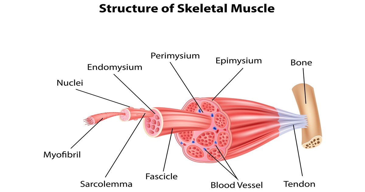
The anatomy of muscle cells differs from that of other body cells and biologists have applied specific terminology to different parts of these cells. Lesson on the anatomy of the forearm: There's no strict demarcation or dividing line between the tendon and the covering around this muscle but that covering is called is called the epimysium fp my cm and it's really just connective tissue that covers the muscle kind of protects it reduces friction. An interactive tutorial teaching the position, actions, innervation and attachments of the rectus femoris muscle with the aid of anatomical illustrations. Muscles of mastication are classified as main and accessory muscles.
Extensor Muscle Anatomy Britannica from cdn.britannica.com Microscopic anatomy of skeletal muscle. Muscle mass accounts for a large majority of the carcass weight of domestic animals. The interactive muscle anatomy diagram shown below outlines the major superficial (i.e. Discover the muscle anatomy of every muscle group in the human body. Practice identifying the major muscles of the human body. Lesson on the anatomy of the forearm: The anatomy of muscle cells differs from that of other body cells and biologists have applied specific terminology to different parts of these cells. Attached to the bones of the skeletal system are about 700 named muscles that make up roughly half of a person's body weight.
Shoulder pain can be a rather complicated matter because of all the image of the muscles shown from a side view is not completely labeled.
They both form the achilles tendon and attach on the posterior aspect of your calcaneus, or heel bone. The muscular system consists of the skeletal muscles and their associated structures. This is a table of skeletal muscles of the human anatomy. The smooth muscle tissue that forms organs like the stomach and bladder changes. Discover the muscle anatomy of every muscle group in the human body. The forearm is the region of the upper limb between the elbow and the wrist. Located immediately below the skin) muscles of the body. Understanding the structure of a muscle fiber. Muscular contraction is necessary for voluntary and involuntary movement of limbs, stabilization of joints, maintaining luminal diameter (in the case of arteries, bowel, etc), and to produce heat. It occupies most of the oral cavity and oropharynx. An interactive tutorial teaching the position, actions, innervation and attachments of the rectus femoris muscle with the aid of anatomical illustrations. The anatomy of muscle cells differs from that of other body cells and biologists have applied specific terminology to different parts of these cells. Muscle mass accounts for a large majority of the carcass weight of domestic animals.
Anatomy of the muscular system. Discover the muscle anatomy of every muscle group in the human body. Smooth muscles (involuntary muscles) are usually in sheets or layers, with one layer of muscle behind the other. The muscles of mastication are a group of muscles associated with movements of the jaw. Muscular contraction is necessary for voluntary and involuntary movement of limbs, stabilization of joints, maintaining luminal diameter (in the case of arteries, bowel, etc), and to produce heat.
Muscle And Tendon Structure Larson Sports And Orthopaedics from larsonsportsortho.com Learn about human anatomy muscles with free interactive flashcards. Cardiac muscle contracts the heart to pump blood. Movement of the mandible at the temporomandibular joint). Attached to the bones of the skeletal system are about 700 named muscles that make up roughly half. Understanding the structure of a muscle fiber. Muscle tendons are extremely important in reinforcing and stabilizing joints. The muscles of mastication are a group of muscles responsible for chewing (i.e. Smooth muscles (involuntary muscles) are usually in sheets or layers, with one layer of muscle behind the other.
There are around 650 skeletal muscles within the typical human body.
Shoulder pain can be a rather complicated matter because of all the image of the muscles shown from a side view is not completely labeled. The muscles around the knee help to keep the knee stable, well aligned, and moving. The muscles of mastication are a group of muscles responsible for chewing (i.e. Muscles of the upper and lower leg. As with muscles of other regions of the body, the various muscles of the upper and lower leg can be divided into groups. Discover the muscle anatomy of every muscle group in the human body. Circular skeletal muscles are made up of fibers explore the minute details of the muscular system in complete anatomy with a suite of 3d learning features such as muscle motion, innervation. They both form the achilles tendon and attach on the posterior aspect of your calcaneus, or heel bone. In the muscular system, muscle tissue is categorized into three distinct types: The muscular system consists of the skeletal muscles and their associated structures. Convergent muscles contain fibers that have a wide origin, but converge in order to attach to a narrow tendon. Each of these muscles is a discrete organ constructed of skeletal muscle tissue, blood vessels, tendons, and nerves. Skeletal muscles are attached to bones by tendons and can be as long as 30 cm, although they are usually 2 to 3 cm in length.
Share :
Post a Comment
for "Anatomy Of Musckes Sndctendons : Muscles Of The Foot Dorsal Plantar Teachmeanatomy"


Post a Comment for "Anatomy Of Musckes Sndctendons : Muscles Of The Foot Dorsal Plantar Teachmeanatomy"